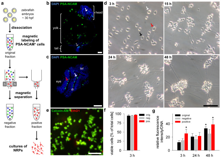
Figure 2. Homogeneous and viable cell cultures of MACS-isolated PSA-NCAM positive cells from embryonic zebrafish.
(a) Schematic representation of magnetic-activated cells sorting (MACS) via magnetic anti-PSA-NCAM microbeads. The single cell suspension of dissociated embryonic zebrafish was incubated with anti-PSA-NCAM microbeads and loaded onto a MACS Column. The magnetically labelled cells were retained in the magnetic field of a MACS Separator, whereas the unlabelled cells flowed through the column (negative fraction). The labelled PSA-NCAM positive cells were then flushed out and collected (positive fraction). (b) Coronal and (c) transversal paraffin sections of embryonic zebrafish (30 hpf) showing PSA-NCAM expression (green) in the tail (grey arrow) as well as in several regions of the brain (white arrows) and the eyes (red arrow). Note the autofluorescence of the yolk. Nuclei were stained with DAPI (blue). tel, telencephalon; di, diencephalon. (d) Primary cultures of adherent cells from the positive fraction (50.000 cells/cm2) showed unipolar (white arrow), bipolar (black arrow) and multipolar (red arrow) morphologies and formed interconnected cell aggregates after 24 h. (e) Viability was assessed by using the Live/Dead assay showing live cells stained with calcein (green) and dead cells with EthD-1 (red). All scale bars, 25 μm. (f) Cells from each fraction showed high viability at 3 h in vitro (n = 3, mean ± SD). (g) Metabolic activity of cells from each fraction was detected by a resazurin-based assay at 3 h, 24 h and 48 h in vitro (n = 5, mean ± SD, P < 0.001). See Fig. 3 for further characterization of PSA-NCAM positive cells and their viability.

