
Figure 3. In vitro cytopathic effects of MV.
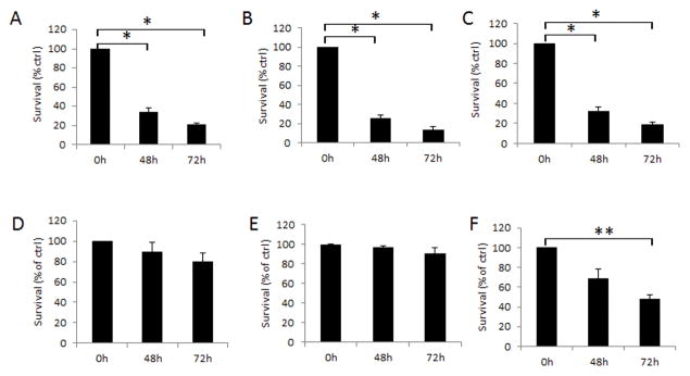
Human breast cancer cells MDA-MB-231 (A), MCF-7 (B), MDA-MB-436 (C) and human mammary epithelial cells HMEC (D) were infected with MV-h-uPA (MOI = 1). Murine mammary epithelial cells MMuMG (E) and mammary cancer cells 4T1 (F) were infected with MV-m-uPA (MOI = 1). Viability was determined at different time points (48h, 72h) by trypan blue exclusion and presented as percentage of controls. Bars represent averages +/− SD of triplicate experiments. * p < 0.0001; ** p = 0.0292.