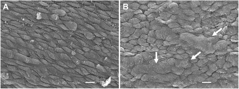
Figure 5.

Scanning electron microscope (SEM) photographs of the vascular endothelium from umbilical arteries of pregnant patients. A) A typical image from a non-smoking patient shows a regular surface accompanying the longitudinal direction of the vessel. B) A typical image from a smoking patient shows diffuse areas of endothelial thickening with loss of the typical architecture and disposition of endothelial cells (white arrows). Scale bar: 10 µm.
