Figure 3.

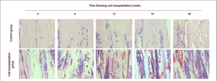
Hematoxylin-eosin staining of rat grafts at 4–20 weeks after cell transplantation (sagittal plane, × 400).
Regenerating nerve fibers are arranged into nerve tracts and abundant capillary vessels are observed.

Hematoxylin-eosin staining of rat grafts at 4–20 weeks after cell transplantation (sagittal plane, × 400).
Regenerating nerve fibers are arranged into nerve tracts and abundant capillary vessels are observed.