
Figure 3.
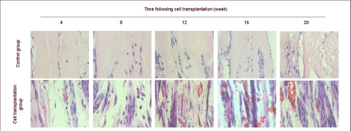
Hematoxylin-eosin staining of rat grafts at 4–20 weeks after cell transplantation (sagittal plane, × 400).
Regenerating nerve fibers are arranged into nerve tracts and abundant capillary vessels are observed.
Official websites use .gov
A
.gov website belongs to an official
government organization in the United States.
Secure .gov websites use HTTPS
A lock (
) or https:// means you've safely
connected to the .gov website. Share sensitive
information only on official, secure websites.
Hematoxylin-eosin staining of rat grafts at 4–20 weeks after cell transplantation (sagittal plane, × 400).
Regenerating nerve fibers are arranged into nerve tracts and abundant capillary vessels are observed.