Figure 2.

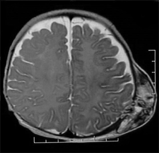
Magnetic resonance imaging axial T2-weighted images showing a heterogeneously hyperintense lesion with multiple flow voids

Magnetic resonance imaging axial T2-weighted images showing a heterogeneously hyperintense lesion with multiple flow voids