Figure 4.

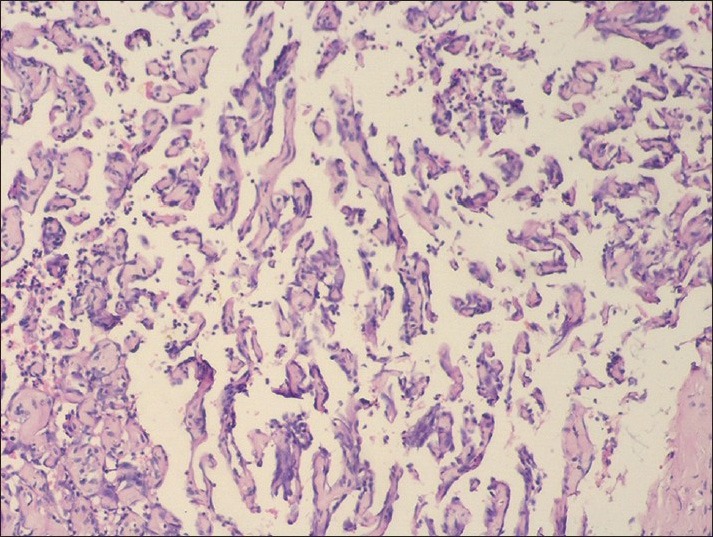
Photomicrograph showing papillary projections within the vascular lumen, having a fibrin core and lined by plump endothelial cells (H and E, ×40)

Photomicrograph showing papillary projections within the vascular lumen, having a fibrin core and lined by plump endothelial cells (H and E, ×40)