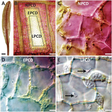
Figure 1.

Developmental PCD during perforation formation in the lace plant. (A) Lace plant with window formation stage leaf. (B) Individual areole between longitudinal and transverse veins showing borders of all three cell types: NPCD, EPCD, and LPCD cells. (C) NPCD cells, pink colouration is due to anthocyanin localized to vacuoles in the underlying mesophyll cells. (D) EPCD cells, showing disappearance of anthocyanin but retention of chloroplasts. (E) LPCD cells, with few remaining chloroplasts. Large aggregates are present in the central vacuole (black arrows). Scale bars: A = 3 mm, B = 150 μm, C-E = 15 μm.
