
Figure 2.
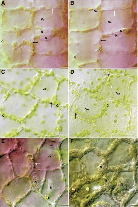
NPCD cells after 10 min heat shock treatments. (A) Cells at 0 h after 45°C treatment. (B) Cells at 6 h after 45°C treatment. Note that tonoplast and PM appears intact. (C) Cells at 0 h after 55°C treatment. Anthocyanin disappearance is evident. (D) Cells at 6 h after 55°C heat shock treatment. Vesicle formation appears throughout cells. (E) NPCD cells before 65°C treatment. (F) NPCD cells after 65°C treatment with disapearance of anthocyanin from the central vacuole. Cellular debris at the periphery of the cells had taken has a textured appearance. (A-D) N– nucleus, C– chloroplast, Va– vacuole, Ve– vesicle, white arrow– tonoplast, black arrow– PM. Scale bars: A-D = 15 μm.