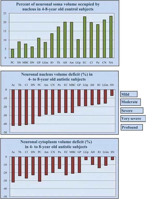
Figure 1.

Region-specific proportion between nucleus and neuron soma volume and the range of neuronal nucleus and cytoplasm volume deficit in children diagnosed with autism. Percent of cell volume occupied by nucleus in 4- to 8-year-old control subjects illustrates neuron type– or region-specific proportions between cell and nucleus volume, with the smallest contribution of nucleus to neuron volume in the largest neurons (Purkinje cell, 5%), and the largest contribution in the smallest neurons (nucleus accumbens, 24%). Neuronal nucleus volume deficits (control 100%) in 4- to 8-year old autistic subjects range from mild (<10%) in the substantia nigra to profound (>40%) in the n. accumbens, thalamus, and claustrum. The cytoplasm volume deficit is less prominent and ranges from 4% in the substantia nigra to 32% in the nucleus accumbens. PC, Purkinje cell; SN, substantia nigra; MBC, magnocellular basal complex; DN, dentate nucleus; GP, globus pallidus; LGNm, magnocellular layer of the lateral geniculate nucleus; IO, inferior olive; Th, thalamus; AH, Ammon’s horn; Am, amygdala; LGNp, parvocellular layer of the lateral geniculate nucleus; EC, entorhinal cortex; Cl, Claustrum; Pu, putamen; CN, caudate nucleus; Ac, nucleus accumbens.
