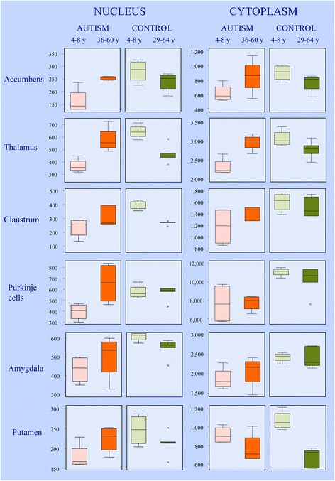
Figure 5.

Difference between neuronal nucleus and cytoplasm volume distribution in control and autism group. Box plot illustrates neuronal nucleus volume distributions in the six brain structures with the most severe volume deficit. The upper and lower boundaries of each box represent the 75th and 25th percentiles of nucleus volume (μm3), respectively. The depth of the box represents the interquartile range (IQR). The whisker above the box marks the maximum value unless any data point lies more than 1.5 times of the IQR above the 75th percentile. The points outside it (outliers) are indicated by circles. The lower whisker and outliers are shown analogously. In all six brain structures, the median volumes of neuronal nuclei in autistic children were below the range of the median volumes of four 4- to 8-year-old control children. The dominant feature in 10 autistic subjects 36–60 years of age was an increase in the volume of nuclei in comparison to in autistic children, as shown as the IQR of older subjects above the median value of children. The level of 75th percentile and the whisker above the box indicate that in the older cohort, the volume of nuclei of the majority of subjects increases with age. The persistence of the volume of neuronal nuclei in the amygdala in a minority of adults at the level of that of 4- to 8-year-old children suggests heterogeneity within the autistic group and indicates a lack of neuronal nucleus increase in some autistic adults. The opposite pattern is observed in the 29- to 64-year-old control group, reflected in reduction of median neuronal nucleus volume and IQR shift below the median value of control 4- to 8-year-old children. In general, the volume of cytoplasm increases with age in adult autistic subjects and decreases in the control group.
