
Figure 3.
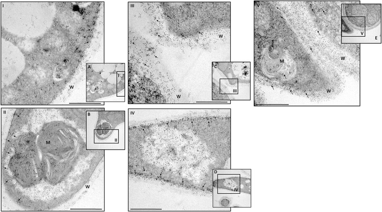
IEM detection of enolase in P. lutzii yeast cells without Cu (A), and without Cu add laminin (B), fibronectin (C), type I collagen (D), or type IV collagen (E). The arrows indicate enolase labeled with gold particles. Bars: 0.07 μm. W: cell wall; M: mitochondria; V: intracellular vesicles. I, II, III, IV and V indicate which image region has been increased in the microscope.