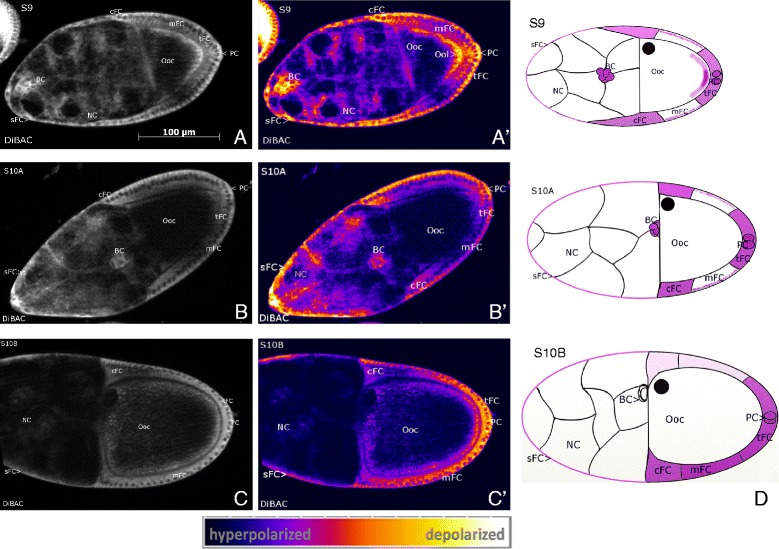
Figure 2.

Characteristic patterns of membrane potentials (V mem ). DiBAC staining, SIM, S9 to S10B, A, B, C: representative grey-scale images, A´, B´, C´: corresponding pseudocolour images. Depolarization is indicated by strong, hyperpolarization by weak fluorescence intensity (see scale bar). D: schematic drawings of characteristic Vmem-patterns; depolarized regions are shown in lilac (staining in NC is not considered). In S9 (A, A´) the FC epithelium is patterned along the anteroposterior axis. Strongest depolarization is found in cFC, sFC, tFC, BC and PC. The mFC are characterized by a hyperpolarized Vmem and an intracellular apicobasal gradient (apical region near Ooc is hyperpolarized compared to basal region), while the anterior region of the Ooc is hyperpolarized relative to the posterior region. In S10B (C, C´) the ventral side of the follicle is depolarized (arrow) in relation to the other side. S10A (B, B´) shows characteristics of both S9 and S10B. For abbreviations, see Figure 1.
