
Fig. 2.
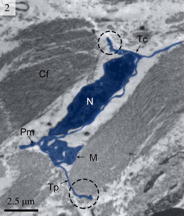
Digitally coloured TEM image of the turtle uterus (enlarged rectangular area of Fig. 1) showing a telocyte with its long telopodes. Circular areas show the close connection of telopodes with collagen fibres. The telopode is very long with alternating podom and podomer. The mitochondria are also visible. (Tc, telocyte; Tp, telopode; Pd, podomer; N, nucleus; M, mitochondria; Cf, collagen fibres). The scale bar represents 2.5 μm.