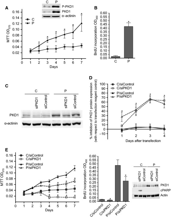
Fig. 1.

PKD1 induces MCF-7 cell proliferation and survival in estrogen-free medium. (A) PKD1-overexpressing (P) and control (C) cells were incubated for different periods of time (1–7 days) in estrogen-free medium. Viable cells were identified over seven days by MTT assay. The results presented are the means ± SD for four independent experiments. *P < 0.01 versus control cells. Inset: immunodetection of phospho-PKD1, PKD1 and α-actinin in cell lysates from C and P cells cultured in estrogen-free medium. (B) Control (C) and PKD1-overexpressing (P) MCF-7 cells were incubated for 48 hrs in estrogen-free medium before BrdU incorporation analysis. The results presented are the means ± SD for two independent experiments. *P < 0.01 versus control cells. (C and D) Control (C) and PKD1-overexpressing (P) MCF-7 cells were transfected with siPKD1 or siControl and cultured in phenol red-free DMEM containing 10% charcoal-treated FBS. From one to four days after transfection, proteins from transfected cells were subjected to SDS-PAGE, transferred to nitrocellulose and immunodetected with anti-PKD1 or anti-α-actinin antibodies. (C) The autoradiograms presented are those of typical experiments performed 4 days after transfection. (D) The percentage of inhibition of PKD1 expression was determined over 4 days after transfection. The results presented are the means ± SD for three independent experiments. *P < 0.01 versus transfection reagent treated cells. (E) PKD1-overexpressing (P) and control (C) cells were transfected with siPKD1 or siControl. The next day, cells were cultured in estrogen-free medium for different periods of time and then analysed as in panels A and B by MTT and BrdU incorporation assays. The results presented are the means ± SEM or SD for three or two independent experiments, respectively. *P < 0.01 versus transfection reagent treated cells. Inset: immunodetection of PKD1, cleaved PARP (cPARP) and actin in cell lysates from C and P cells transfected with siControl or siPKD1 and cultured for 3 days in estrogen-free medium.
