
Figure 5.
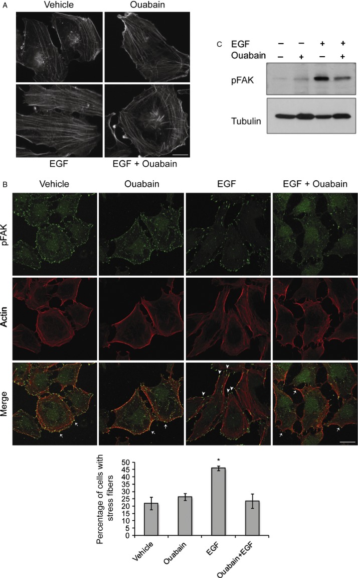
Ouabain prevents EGF-induced stress fiber formation. (A) Epifluorescence images of actin stress fibers in control cells and DAOY cells treated with ouabain, EGF, and EGF + ouabain for 2 h after FITC-conjugated phalloidin staining. Bar, 12.5 μm. (B) Confocal images of activated, phosphorylated FAK (green), and actin stress fibers (red) in DAOY cells treated with and without EGF and ouabain as in (A). Arrows mark lamellipodia and arrowheads indicate phospho-FAK localized to the tips of actin stress fibers. Bar, 25 μm. Graph shows quantitative data of number of cells with stress fibers relative to total number of cells counted per treatment group and represent average ± SE of three independent experiments, *P < 0.001. (C) DAOY cells were incubated with 10 ng/mL EGF in the presence or absence of 50 μmol/L ouabain. Activation of FAK was monitored by immunoblotting with phospho-specific anti-FAK antibody. Tubulin immunoblot confirms equal loading. EGF, epidermal growth factor; FAK, focal adhesion kinase.