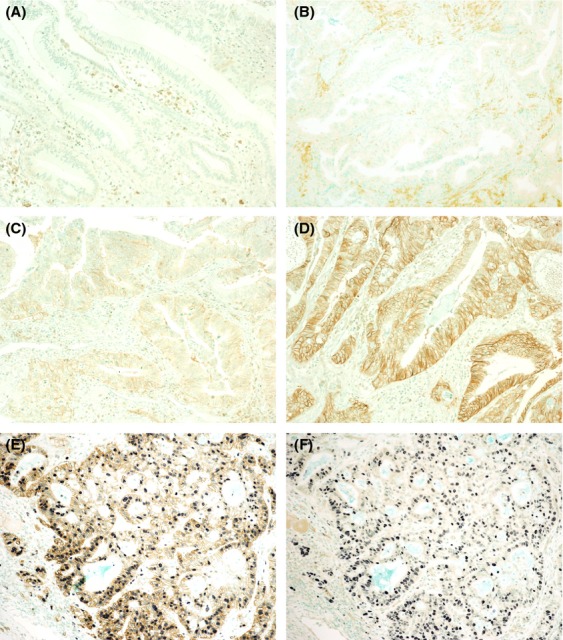
Figure 1.

L-type amino acid transporter (LAT) 1 expression in bile duct carcinoma (BDC) cells analyzed by immunohistochemistry. Based on the immunointensity of the carcinoma cell membrane, four categories were defined: A, 0, no staining; B, 1, weak or patchily positive staining; C, 2, moderate cell membrane staining; and D, 3, intense complete membrane staining. Activated lymphocytes also showed LAT1 expression. E and F, double staining for LAT1 (E) or LAT2 (F) and Ki-67 in a representative patient with BDC. Many carcinoma cells that express Ki-67 (nuclear staining with NiCl2-DAB, blue) coexpress LAT1 (membranous staining with DAB, brown), but not LAT2. Slides were counter-stained with methyl green solution. Original magnification, ×100 (A through D), ×200 (E, F).
