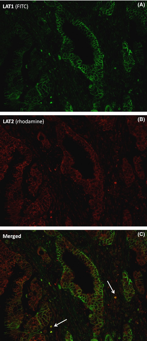
Figure 3.

Double immunofluorescence staining with LAT1 and LAT2. Bile duct carcinoma (BDC) cells did not coexpress LAT1 (fluorescein isothiocyanate, green) and LAT2 (rhodamine, red), although some activated lymphocytes (arrow) showed both LAT1 and LAT2 expression (merged, yellow). Original magnification, ×200.
