
Figure 4.
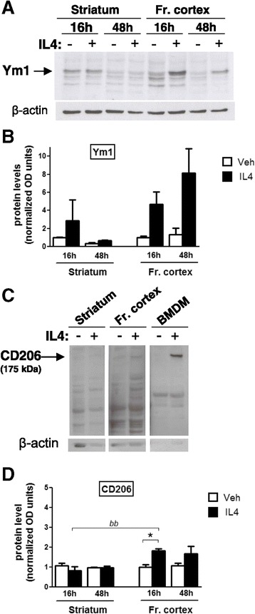
Time course of IL4-induced M2a protein expression in mouse brain. After intracerebroventricular injection of saline (Veh, open bars) or IL4 (black bars), the striatum and frontal cortex (Fr. cortex) were extracted at 16 and 48 h intervals and analyzed by Western blotting to evaluate Ym1 (A,B) and CD206 (C,D) protein levels. (A,C) Immunoblots using Ym1, CD206, and β-actin antibodies from representative samples are shown. The average densitometric value of the Ym1 (B) and CD206 (D) bands from Western blots of several samples was normalized to that of β-actin; bars represent mean values ± SEM; the arbitrary value of 1 was given to the value of the saline 16 h group for each protein and brain area analyzed (n = 4 to 6). BMDM, bone-marrow-derived macrophages.