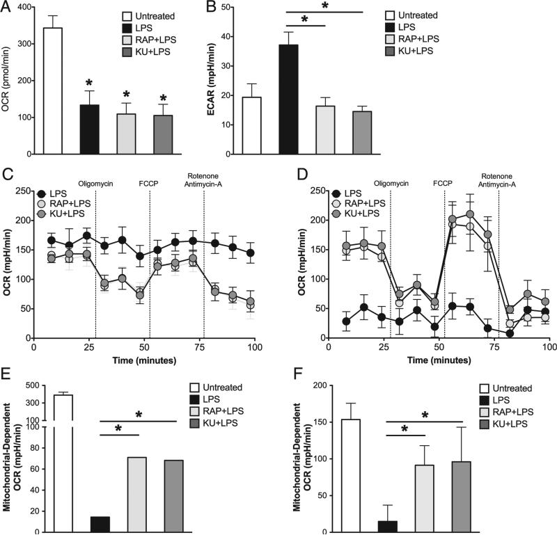
FIGURE 4.
Mitochondrial OXPHOS is preserved in GM-DCs activated by LPS in the presence of mTOR inhibitors. (A and B) GM-DCs were either left unstimulated or activated with LPS for 24 h in the presence or absence of RAP or KU. Basal oxygen OCRs (A) and ECARs (B) for each treatment group were determined by an XF extracellular flux analyzer assay. (C and D) GM-DCs were stimulated with LPS in the presence or absence of RAP or KU for 24 (C) and 72 h (D). Mitochondrial function was assessed by an XF extracellular flux analyzer assay. (E and F) Mitochondrial-dependent OCR was determined for experiments described in (C) and (D) by subtracting residual OCR after antimycin A/rotenone treatment from basal OCR levels. This calculation was performed for GM-DCs after 24 h stimulation (E) and 72 h after stimulation (F). Graphs in this figure represent mean values ± SD of at three independent experiments. *p < 0.05.

