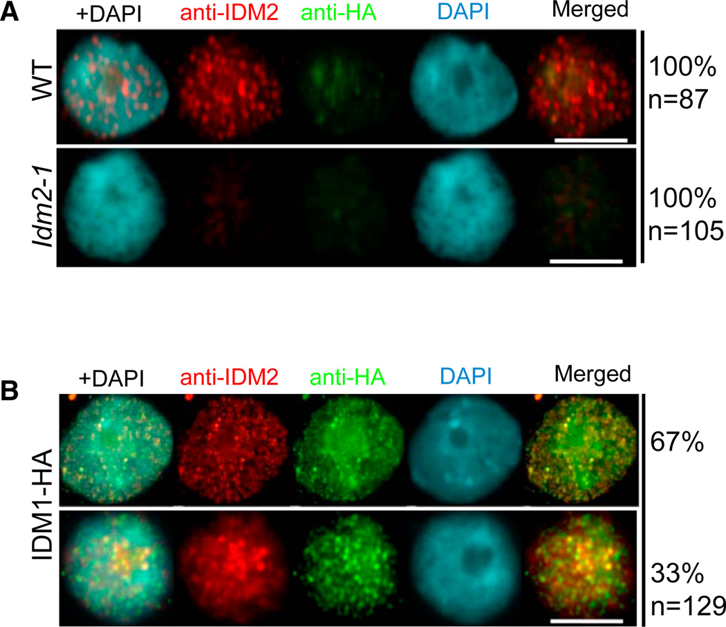
Figure 5. Subnuclear Localization of IDM2 and Its Colocalization with IDM1.
(A) Detection of IDM2 (red) in the wild-type and idm2-1 mutant nuclei by immunostaining using anti-iDM2. Scale bars represent 10 µm.
(B) Dual immunolocalization of IDM2 (red) and IDM1-HA (green). DNA was stained with DAPI (blue). The frequency of nuclei displaying each interphase pattern is shown on the right. Scale bars represent 10 µm. See also Figure S5.

