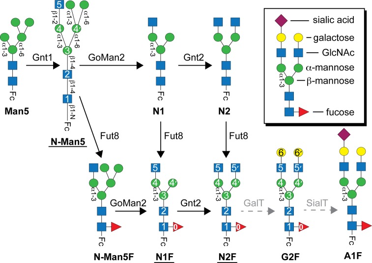
Figure 1.
Native IgG1 Fc N-glycan processing in the Golgi. Conversions catalyzed by the enzymes indicated above the solid arrows and labeled with black type largely proceed to completion. Reactions catalyzed by enzymes denoted with gray type a dashed arrow modify some but not all of the secreted IgG1. Glycoforms studied here by nuclear magnetic resonance are underlined. Carbohydrate residues are numbered according to ref (30) and represented using the CFG convention and shown in the inset49 (GlcNAc, N-acetylglucosamine). Glycosidic linkages of the human IgG1 Fc N-glycan are indicated.

