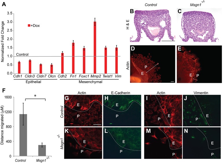
Fig. 6.
Msgn1 is required for EMT and motility. (A) qPCR analysis of epithelial and mesenchymal genes expressed in Dox-treated (+Dox) iF-Msgn1 EBs after 48 h. The y-axis shows the normalized fold change; error bars are the s.d. (B,C) Histological analysis (H&E) of PS cross-sections of control (n=7) and Msgn1−/− (n=9) embryos at E8.5. (D,E) Rhodamine-phalloidin staining of control (D, n=21) and Msgn1−/− (E, n=14) tail bud explants dissected at the 18-22 somite stage and cultured for 48 h. The distance migrated from explant (E) to periphery (P) is indicated by white line. (F) The average distance migrated in control (n=13) and Msgn1−/− (n=7) explants is shown on y-axis. *P≤9.88×10⁻⁵, two-sample unequal variance Student's t-test. (G-N) Immunostaining of control and Msgn1−/− explants cultured for 48 h and stained with Rhodamine-phalloidin, and anti-E-cadherin and anti-vimentin antibodies. The dashed white line demarcates the explant (E) from periphery (P) region. Scale bars: 100 μm.

