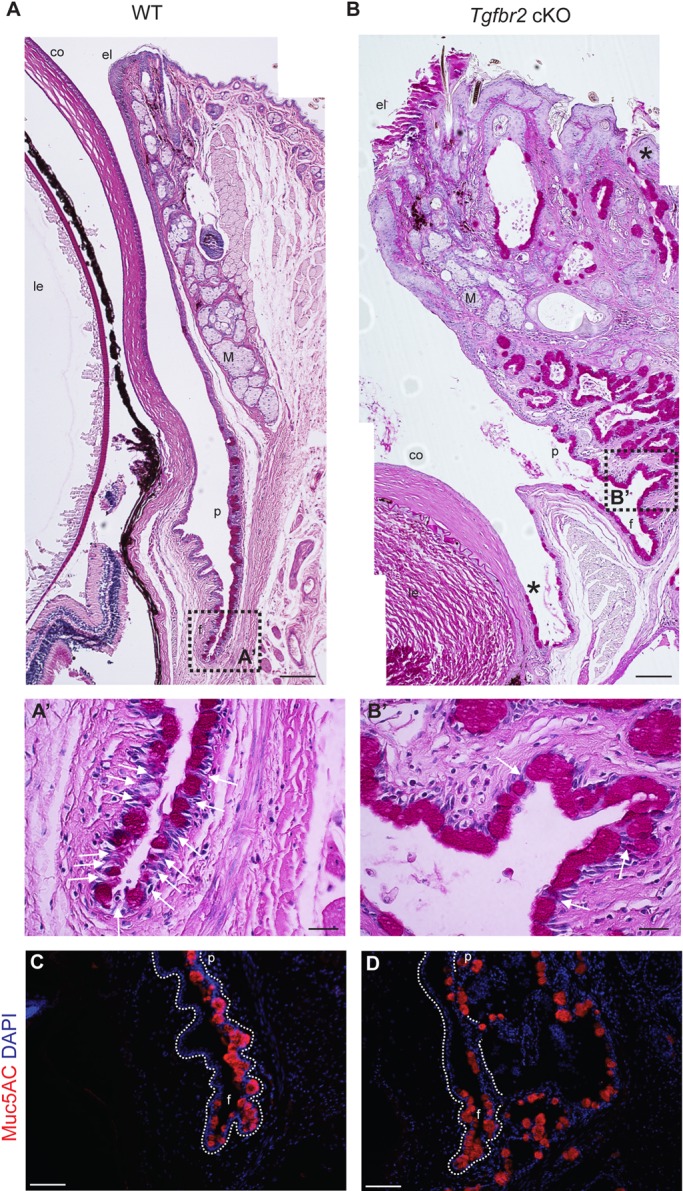
Fig. 2.

Tgfbr2-deficient mice develop eyelid, corneal and conjunctival epithelial hyperplasia. (A-B′) Combined PAS and Hematoxylin and Eosin staining demonstrated extensive squamous and mucous epithelial hyperplasia with invaginations into the underlying stroma, involving the palpebral conjunctiva, fornix and eyelids of symptomatic Tgfbr2 cKO mice compared with sections of comparable regions from age-matched wild-type mice. Ectopic goblet cells (magenta) were found within Tgfbr2 cKO hyperplastic eyelid epithelium and peripheral corneal epithelium (B, asterisks). Higher magnification of the boxed areas are shown in A′ and B′. White arrows indicate non-goblet cell stratified conjunctival epithelial cells interspersed between goblet cells. (C,D) Goblet cells within the expanded and invaginated Tgfbr2 cKO conjunctiva expressed Muc5AC. Dotted lines indicate the basal layer. DAPI counterstains nuclei in blue. co, cornea; le, lens; f, fornix; p, palpebral conjunctiva; M, Meibomian gland; el, eyelid; f, fornix. Scale bars: 100 µm in A,B,C,D; 20 µm in A′,B′.
