
Fig. 5.
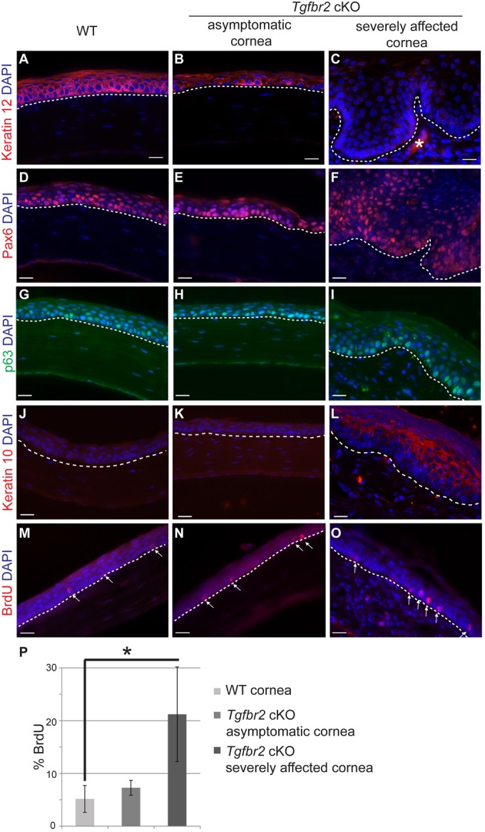
The hyperplastic Tgfbr2-deficient adult cornea becomes keratinized. (A-C) Immunofluorescence staining with antibodies against keratin 12 (A-C), Pax6 (D-F), p63 (G-I), keratin 10 (J-L) and BrdU (M-O) indicated that the corneal epithelium of adult Tgfbr2 cKO mice becomes keratinized with increasing phenotypic severity. (M-P) Quantification (P) of BrdU immunofluorescence staining (M-O, arrows) indicated that the severely affected Tgfbr2 cKO corneal epithelium was hyperproliferative compared with wild-type and asymptomatic Tgfbr2 cKO adult corneal epithelium. Data represent the mean number of BrdU-positive cells in relation to total basal epithelial cells ±s.d.; Student's t-test; *P=0.04. Dotted lines indicate the basal layer. Asterisk in C denotes autofluorescence in the stroma. DAPI counterstains nuclei in blue. Scale bars: 20 µm.