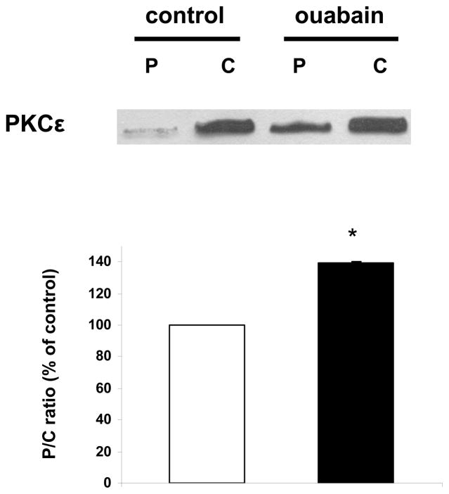
Figure 4. Effect of ouabain on PKCε activation.
Rabbit hearts were perfused for 20 min in the presence or absence of ouabain 500 nM (protocol A). Cytosolic (C) and particulate (P) fractions obtained from tissue lysates were then assayed and compared for their contents in PKCε. Upper panel: representative western blot. Lower panel: means ± SEM of 3 separate experiments. * P < 0.01 vs control.

