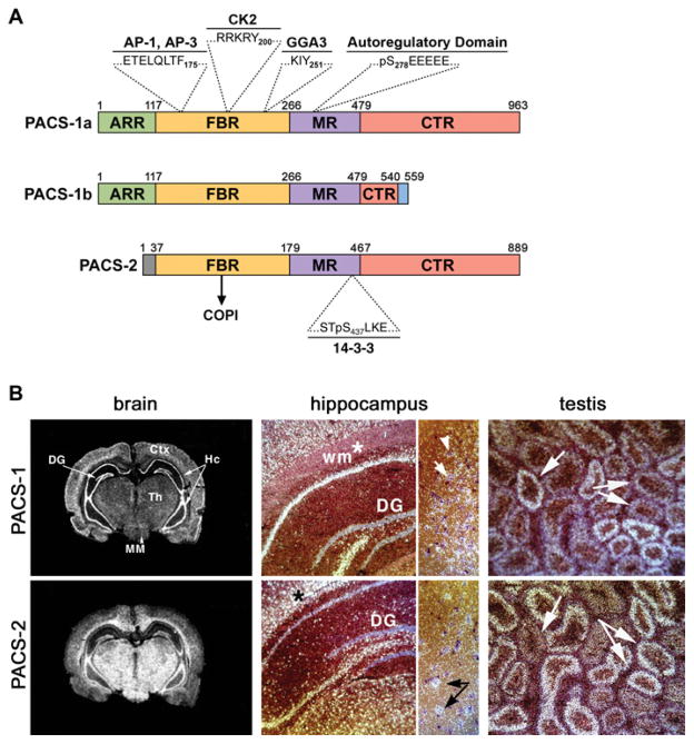
Figure 1. Domain organization of the PACS proteins.
(A) Schematic of PACS-1 and PACS-2 illustrating the proposed domains and residues important for partner protein binding. (B) In situ hybridization of PACS-1 and PACS-2. Left-hand panels, coronal sections of rat brain stained with PACS-1 or PACS-2 cRNA probes. DG, dentate gyrus; Hc, hippocampus; Ctx, cortex; Th, thalamus; MM, medial mammillary nucleus. Middle panels, darkfield staining of hippocampus showing neuronal and glial labelling. (*), alignment marker; white arrows, neurons; black arrows, glia. Right-hand panels, PACS-1 and PACS-2 in consecutive serial sections of the testis. Arrows mark seminiferous tubules with inverse staining of PACS-1 and PACS-2. Sense probes showed no staining.

