Figure 3.

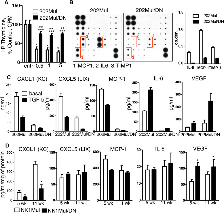
Cytokine and chemokine profile of Erbb2+ mammary carcinoma cells with intact and modified transforming growth factor β (TGFβ) signaling. (A) Thymidine incorporation assay. Epithelial cells were treated with 0.5, 1 and 5 ng/mL TGFβ and analyzed for changes in cell proliferation by 3(H) thymidine incorporation. Statistical significance was determined by P-values <0.05; *202Mul treated versus 202Mul untreated, **202Mul/DN treated versus 202Mul/DN untreated, ***202Mul/DN treated versus 202Mul treated. (B) Representative mouse cytokine array using tumor explant supernatants [45] from 202Mul and 202Mul/DN cell lines (left) quantification of the mouse cytokine array data for IL-6, monocyte chemotactic protein 1 (MCP1) and tissue inhibitor of metalloproteinase 1 (TIMP1) (right). (C) ELISA of conditional medium from 202Mul and 202Mul/DN cell lines untreated and treated for 24 hours with TGFβ 1 ng/mL. All changes in cytokine and chemokine secretion were significant (P <0.05) in 202Mul versus 202Mul/DN cell lines and treated versus untreated with TGFβ. (D) ELISA of protein extracts from tumor tissue homogenates isolated from NK1Mul and NK1Mul/DN mice on 5 and 11 weeks after tumor palpation. Five mice per group were used excluding vascular endothelial growth factor (VEGF) measurement, where eight mice were used; *P <0.05. Data correspond to the mean ± standard error of the mean.
