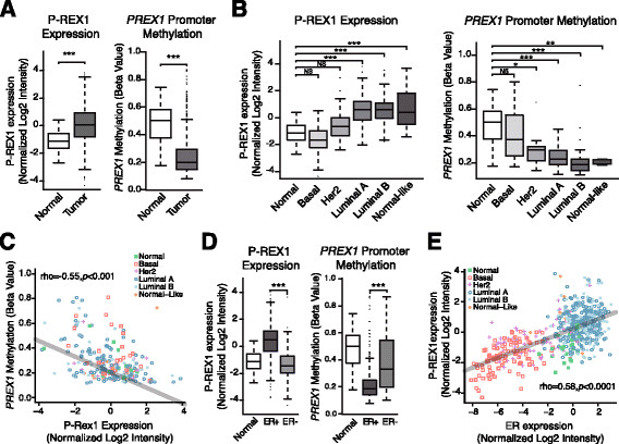
Figure 4.

Analysis of PREX1 methylation and mRNA expression in the TCGA dataset. Panel A . P-REX1 mRNA expression (left) and promoter methylation (right) in breast tumors and normal breast tissue (*** = P <0.0001). Panel B . P-REX1 mRNA expression (left) and promoter methylation (right) across different breast cancer subtypes (*, P <0.05; **, P <0.01; ***, P <0.001). Panel C . Inverse correlation between P-REX1 mRNA levels and PREX1 methylation in different subtypes of breast tumors and normal breast tissue (Spearman rho = -0.55, P <0.0001). Panel D . P-REX1 mRNA expression (left) and promoter methylation (right) in normal tissue, estrogen receptor (ER)-positive and ER-negative breast tumors (***, P <0.0001). Panel E . Correlation analysis between P-REX1 and ER-alpha expression in different subtypes of human breast tumors and normal breast samples (Spearman rho =0.85, P <0.0001). TCGA, The Cancer Genome Atlas.
