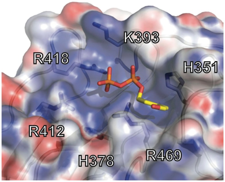
Figure 7.
Phosphoantigen binding pocket of B30.2 domain. Close-up view of the B30.2 pAg binding pocket with the side chains lining the pocket shown under the semi-transparent surface. The positions are labeled with the numbering relative to the full-length BTN3A1 molecule. The pAg is shown as sticks, modeled into the binding pocket; phosphates are colored orange and red (oxygen) and the organic moiety is shown in yellow.

