
Figure 2.
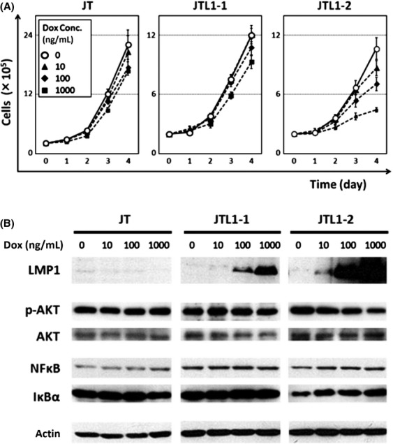
Cell proliferation and levels of signaling molecules in JT, JTL1-1, and JTL1-2 cells. (A) Cell proliferation was assessed by trypan blue staining, followed by cell counting, at days 0, 1, 2, 3, and 4 after the induction of LMP1 with 0, 10, 100, or 1000 ng/mL Dox. Experiments were performed in triplicate, and standard errors and means are shown. (B) Cell extracts harvested 2 days after Dox induction (0, 10, 100, 1000 ng/mL) were analyzed by western blotting. AKT and NFκB signalings were assessed by AKT phosphorylation and the expression of NFκB (p65) and IκBα, respectively.