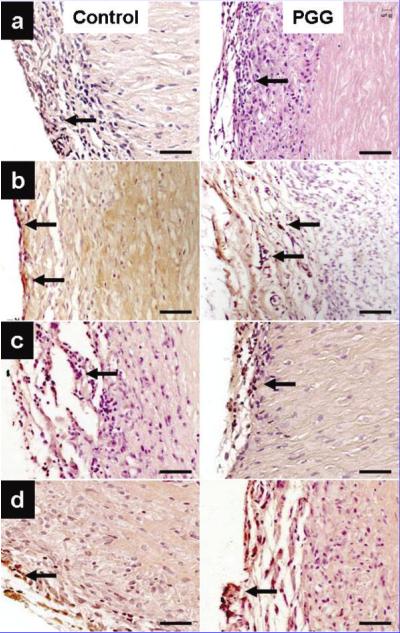
Figure 3.
Identification of infiltrating cells in implanted elastin scaffolds. PGG-stabilized scaffolds were compared to untreated control scaffolds. (a) Vimentin stain showing small numbers of fibroblast-like cells. (b) Prolyl-4-hydroxylase immunohisto-chemical staining: arrows point to positively stained cells. (c) IHC staining showing few infiltrating macrophages (arrows). (d) Alpha-smooth muscle cell staining identifying cells at the periphery of the implants. Sections were counterstained with hematoxylin (nuclei dark blue). Bars are 50 μm in all micrographs.

