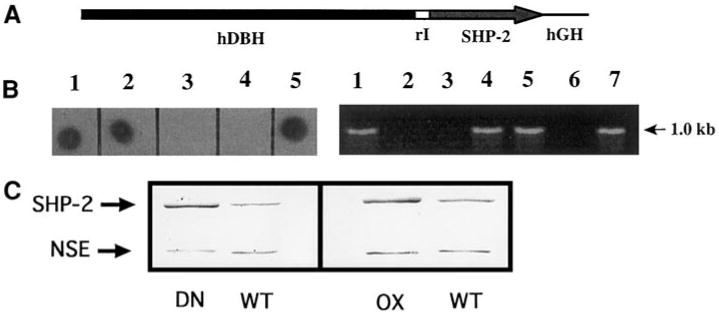
Fig. 1.

Production of SHP-2 transgenic mice. (A) DNA construct used to generate SHP-2 transgenic mice. A 5.8-kb regulatory sequence from the human dopamine β-hydroxylase gene (hDBH) drives expression of a 1.9-kb SHP-2 wild type or Cys→Ser mutant cDNA. rI, 171-bp segment spanning the first intron of rat insulin II gene; hGH, 799-bp portion of the human growth hormone gene. (B) The SHP-2 transgene detected by dot blot of mouse tail DNA with the hGH sequences or by genomic PCR using a primer pair corresponding to the SHP-2 cDNA and the hGH tag, respectively. (Left) 3/5 mice in a litter carry the transgene (lanes 1, 2, 5). (Right) 4/7 mice in a litter were identified as transgenic by genomic PCR (lanes 1, 4, 5, 7). Lanes 3, 4 (left) and 2, 3, 6 (right) contain DNA from wt littermates. (C) SHP-2 protein is overexpressed in superior cervical ganglia (SCGs) from transgenic animals. (Left) 8 μg of protein from a pair of wt or DN SCGs was run on SDS PAGE and detected on a Western blot with anti-SHP-2 antibody. (Right) 5 μg of protein from a pair of wt or OX SCGs was run on SDS PAGE and detected on a Western blot with anti-SHP-2 antibody. After quantification of these bands, blots was reprobed with anti-neuron-specific enolase (NSE) for normalization. In each case, quantitative analysis showed that the transgenic ganglia had a SHP-2/NSE ratio of roughly 2.5. Quantification of blots (n ≥ 3 for each line), using both this normalization method and the slope method (see Materials and Methods) showed that SCGs from both DN and OX lines consistently contained 2-2.5 times normal levels of immunoreactive SHP-2.
