
Fig. 3.
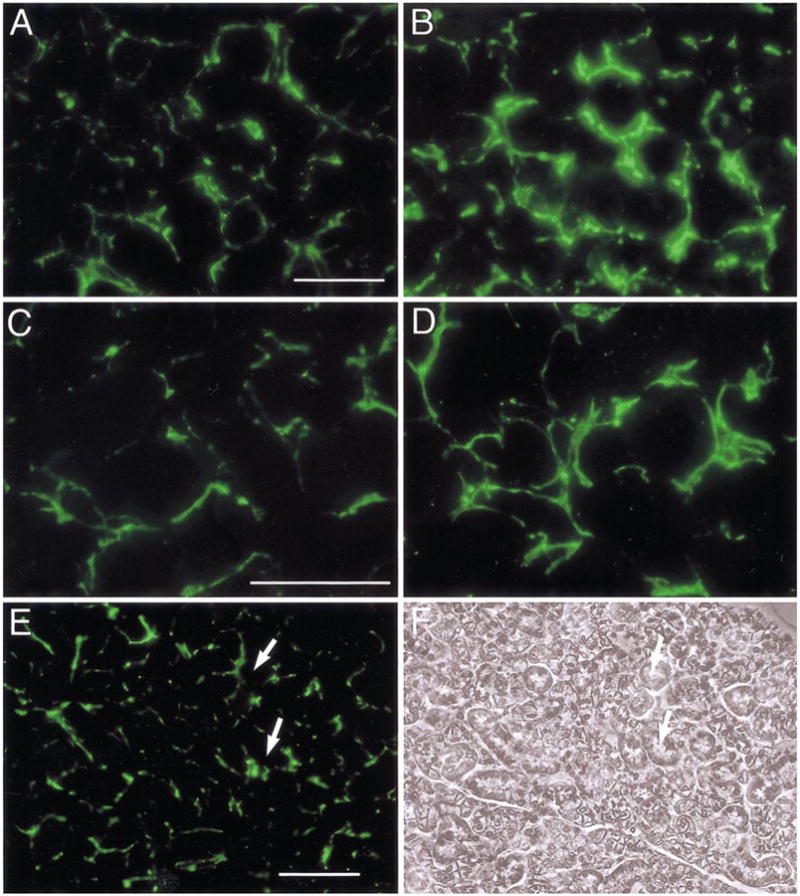
Increased sympathetic innervation of submaxillary glands in SHP-2 DN mice. Sympathetic innervation of submaxillary glands was examined by using antibody to tyrosine hydroxlase (TH), detected with secondary antibodies labeled with Alexa488. (A) In wt mice, individual acini from submaxillary glands are surrounded by TH-positive nerve fibers. (B) In SHP-2 DN glands, staining is noticeably brighter throughout the tissue. (C, D) At higher magnification, an increased number of TH-positive nerve fibers can be seen in the SHP-DN tissue (D), compared with the wt (C). (E) and (F) are TH immunostaining and phase contrast views, respectively, of a DN SHP-2 section at low power. White arrows point to corresponding points in the two micrographs. Scale bar, 30 μm (A, C); 100 μm (E). In this and subsequent figures showing fluorescent images, different tissues were processed together, photographs were taken at the same time under identical illumination conditions, and these were processed identically to facilitate comparison. Representative sections are shown.