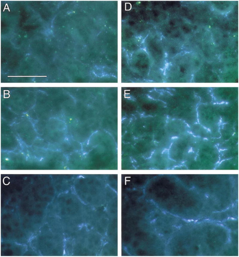
Fig. 4.

Increased sympathetic innervation of submaxillary glands in SHP-2 DN mice, but not in SHP-2 OX mice. Sympathetic innervation of submaxillary glands was examined by using the glyoxylic acid method for catecholamine histofluorescence. (A, D) Wt (A) and SHP-2 DN (D) gland sections in control conditions. There is increased catecholamine fluorescence in the DN tissue. (B, E) Wt (B) and SHP-2 DN (E) gland sections following injection of α-methyl norepinephrine to saturate catecholamine levels. The increased catecholamine fluorescence in the DN is even more evident, suggesting that the increased levels are due to more sympathetic fibers, rather than increased NE levels in the fibers. Examples shown are representative of multiple experiments (n ≥ 4) on each of three SHP-2 DN lines. (C, F) Wt (C) and SHP-2 OX (F) gland sections following injection of α-methyl norepinephrine to saturate catecholamine levels. Levels of catecholamine fluorescence in the OX gland is similar to that seen in the wt gland. Examples shown are representative of multiple experiments (n ≥ 4) on each of 2 SHP-2 OX lines. Scale bar, 30 μm.
