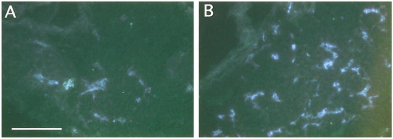
Fig. 5.

Increased sympathetic innervation of pineal glands in SHP-2 DN mice. Sympathetic innervation of pineal glands was examined as for submaxillary glands. Animals were injected intraperitoneally with α-methyl norepinephrine prior to dissection to saturate catecholamine levels. (A) Representative section illustrating fluorescence levels in the wt pineal. (B) Representative section illustrating fluorescence levels in the SHP-2 DN pineal. Fluroescence levels are clearly higher in the SHP-2 DN gland. Similar results were obtained in three independent experiments. Scale bar, 100 μm.
