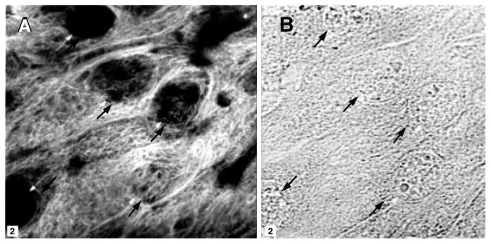
Fig. 1.

Localization of cytokeratin in cultured guinea pig gallbladder cells. A: fluorescence image. B: corresponding bright-field image. The box marked on each image is 2 μm2. The arrows mark the locations of individual nuclei for the alignment of the 2 images.
