
Figure 4. Frames of the Proof-of-Principle Analysis.
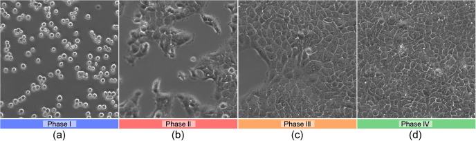
Selected frames of the video in the proof-of-principle analysis. In the first part of the video ((a), phase I), the recorded cells from the epidermoid carcinoma cell line A431 move freely in the nutrition solution until they adhere to the viewing glass. In the second phase, more and more of the visible region is occupied due to cell proliferation ((b), phase II). One part of the recorded area stays blank for a longer time ((c), phase III), before this part is occupied as well and the whole recorded area is occupied by a complete monolayer of the cells ((d), phase IV).