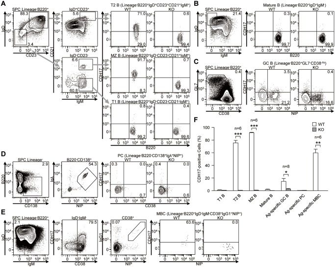
Figure 1. The expression of CDH17 is regulated differentiation-dependently during B cell development in spleen.
(A–E) Expression of CDH17 on spleen cells and bone marrow cells isolated from wild-type (WT) and CDH17-/- (KO) mice were analyzed by flow cytometry: (A) T1 B cells (Lin-B220+IgD-CD23-CD21-IgM+), T2 B cells (Lin-B220+IgD+CD23+CD21+IgM+), and MZ B cells (Lin-B220+IgD-CD23-CD21+IgM+); (B) Mature B cells (Lin-B220+IgD+IgM-); (C) Antigen-specific GC B cells (Lin-B220+GL7+CD38low/-NIP+); (D) Antigen-specific PCs (Lin-B220-CD138+Igλ+NIP+); (E) Antigen-specific MBCs (Lin-B220+IgD-IgM-CD38+IgG1+NIP+). Numbers adjacent to the gates indicate the percentage (%) of cells in the respective parental gates. (F) The percentages of CDH17+ cells within various B cell populations are plotted on a bar graph. The percentage of CDH17+ cells relative to the total number of cells in the parental gates (indicated on the top of the corresponding flow cytometric contour plot) was calculated. In (C), the percentage of antigen-specific cells within the parental gates was calculated (n = 2 (PCs), n = 8 (GC B), n = 6 (others); *P≤0.05, **P≤0.01, ***P≤0.001 (Student’s t-test)).

