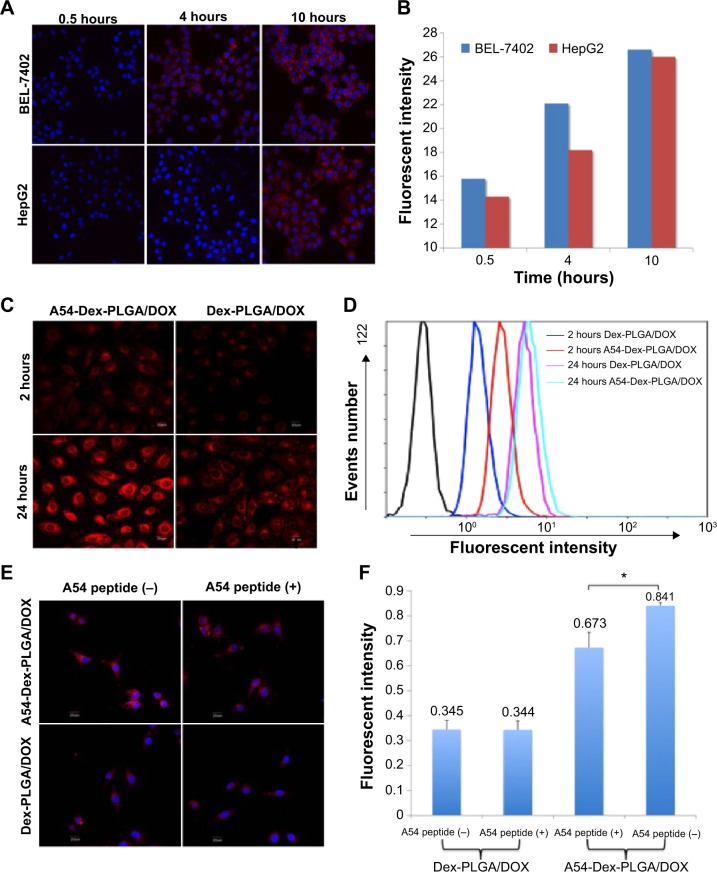
Figure 5.
Cellular uptake results of DOX-loaded micelles.
Notes: Fluorescence images (A) of DOX after BEL-7402 and HepG2 cells were incubated with A54-Dex-PLGA/DOX micelles for 0.5, 4, and 10 hours. Quantitative analysis results (B) based on images (A) by ImageJ software. Fluorescence images (C) after BEL-7402 cells were incubated with DOX-loaded micelles solution for 2 and 24 hours. Quantitative cellular uptake (D) analyzed based on images (C) by a flow cytometry. Fluorescence images (E) of DOX after BEL-7402 was incubated with Dex-PLGA/DOX micelles, A54-Dex-PLGA/DOX micelles, and their blocking ones for 4 hours. Quantitative cellular uptake (F) analyzed based on images (E) by a flow cytometry (*P<0.05).
Abbreviation: A54-Dex-PLGA/DOX, doxorubicin-loaded A54 peptide-functionalized poly(lactic-co-glycolic acid)-grafted dextran.

