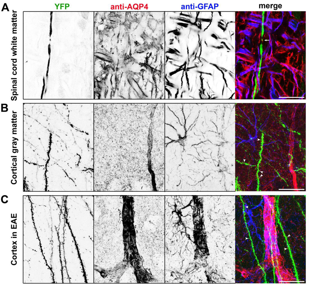
Figure 1. Astrocytes in neurovascular units in white and gray matter.
(A) Axons (YFP in green in merged), astrocytes (GFAP staining in blue in merged) and astrocytic endfeet (AQP4 staining in red in merged) in spinal cord white matter. The white arrow, a putative node of Ranvier. (B) Neurovascular units in cortical gray matter. Axons, dendrites and dendritic spines are in green. White arrowheads, dendritic spines. (C) Neurovascular units in cortical gray matter at the peak stage of EAE (Experimental Autoimmune Encephalomyelitis). These sections were from Thy1:YFP transgenic mice. Confocal image stacks (40 µm in thickness) were collapsed into 2D images. These images are modified from our recent paper [159].

