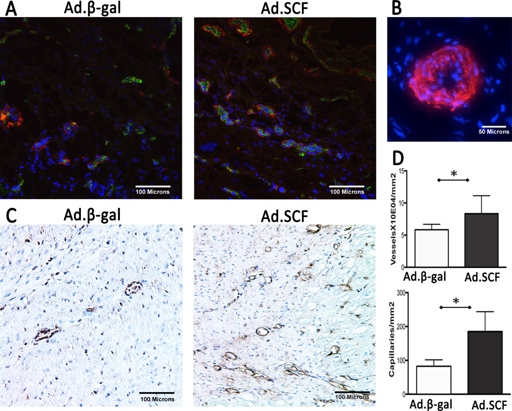
Figure 5. Angiogenesis and vasculogenesis at 3 months after myocardial infarction.
A. Increased numbers of vasculatures co-stained with α-SMA and CD31 were found after Ad.SCF injection compared to the Ad.β-gal. Blue; DAPI-stained nuclei, red; α-SMA, green; CD31. B. Presumably functional vessel stained with α-SMA. Multiple layers of nuclei are found in the vessel wall suggesting that this is a functional arteriole. C. A significant increase in capillaries co-expressing isolectin IB4 (brown) was found at the infarct border after SCF gene transfer. D. Quantification of vessels (P=0.01) and capillaries (P=0.003) suggests angiogenic role of SCF.
*; P<0.05 Abbreviations: α-SMA = α-smooth muscle actin, SCF = stem cell factor

