
Figure 3.
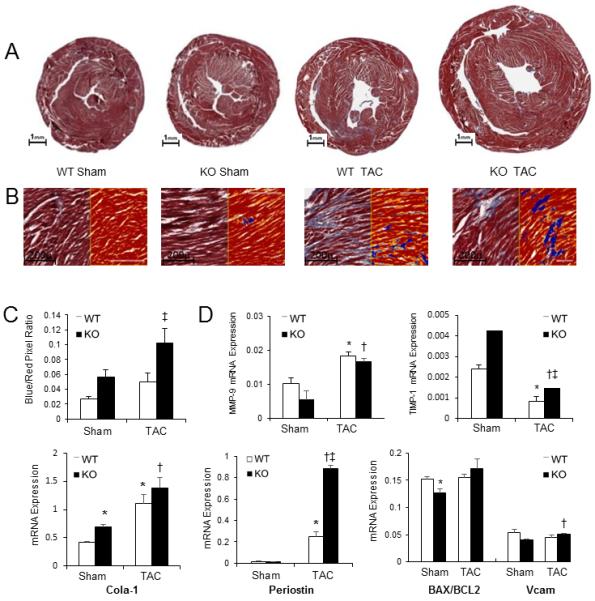
Influence of absence of cardiomyocyte p65 NF-κB on myocardial fibrosis 6-weeks following TAC. (A) Mid-papillary cross-section of mouse hearts stained with Masson’s trichrome. (B) Positive pixel count digital analysis for fibrosis quantification (20x magnification). Blue represents collagen fibers; red represents cardiomyocytes. (C) Quantification of fibrosis as assessed by pixel count ratio. (D) Real-time PCR for fibrotic markers matrix metallopeptidase 9 (MMP9), tissue inhibitor of metallopeptidase 1 (TIMP1), collagen 1α1 (Col1α1), vascular cell adhesion molecule (VCAM), Bax/Bcl2, and periostin. (p<0.05, *vs. WT sham, †vs. KO sham, ‡vs. WT post-TAC).