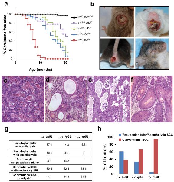
Figure 1. Cooperation of loss of p53 and αv integrin during SCC development.
(a) Kinetics of SCC development induced by deletion of p53 and αv in stratified epithelia. Percentage of carcinoma-free mice are represented over time in mice with the following genotypes: αvf/f-p53wt/wt (black line; n = 26), αvwt/wt-p53f/f (purple line; n = 16), αvf/wt-p53f/f (green line; n = 20), αvf/f-p53f/wt (blue line; n = 34), αvf/f-p53f/f (red line; n = 39). p< 0.05 for the following comparisons: αvf/f-p53f/f with each of the other groups; αvf/f-p53f/wt with αvf/f-p53wt/wt and αvwt/wt-p53f/f; αvf/wt-p53f/f with αvf/f-p53wt/wt. (b) Gross appearance of tumors that developed in the skin, mouth, anal epithelium and eyelid of αvf/f-p53f/f mice. (c–f) Hematoxylin and eosin staining of the primary phenotypes observed in SCCs that developed in αvf/f-p53f/f mice. (g) Quantification of the main phenotypic variants observed in SCCs that lacked both αv and p53 (αv−/p53−), only αv (αv−/p53+), or only p53 (αv+/p53−). (h) Graphical representation of the relative proportion of pseudoglandular/acantholytic SCCs and conventional SCCs in αv−/p53−, αv−/p53+, and αv+/p53− tumors.

