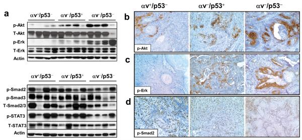
Figure 2. Activation of Akt and Erk in SCCs that lack both p53 and αv.
(a) The top panel shows a western blot analysis with specific antibodies for phosphorylated Akt (p-Akt) and phosphorylated Erk (p-Erk) and the corresponding total proteins (T-Akt and T-Erk, respectively) in representative αv+/p53−, αv−/p53+, and αv−/p53− tumors. The bottom panel shows a western blot for phosphorylated Smad2 (p-Smad2), phosphorylated Smad3 (p-Smad3), and phosphorylated STAT3 (p-STAT3), and the corresponding total proteins (T-Smad2/3 and T-STAT3) using total protein lysates from representative αv+/p53−, αv−/p53+ and αv−/p53− tumors. Actin was used as a loading control. (b-d) Immunohistochemical staining for p-Akt (b), p-Erk (c), and p-Smad2 (d) in the indicated tumors. Note the strong staining for p-Akt and p-Erk in the epithelial component of the αv−/p53− tumors, and p-Smad2 in the nuclei of epithelial cells.

