Figure 1.
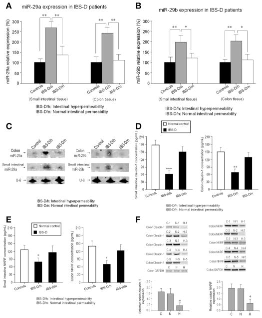
Increased intestinal permeability and miR-29 expression in humans: (A) Real-time polymerase chain reaction assay of miR-29a expression in human small intestine and colon from IBS-D patients. Significant upregulation of miR-29a expression in IBS-D/h patients (n = 40) compared with both controls (n = 36) and IBS-D/n patients (n = 69) (**P < .01) was present in both colon and small intestine. There were no significant differences in miR-29a expression between IBS-D/n patients and controls. (B) Analysis of miR-29b expression in small intestinal and colon tissue showed similar changes as miR-29a. Significant changes in IBS-D/h patients compared to normal controls (**P < .01) and to IBS-D/n patients (*P < .05) were present. (C) Northern blots: Left panel shows miR-29a expression from 3 subjects (control, IBS-D/h, IBS-D/n); right panel shows corresponding blots for miR-29b. These results further confirm the PCR data in (A) and (B). (D) Enzyme-linked immunosorbent assay for CLDN1 expression in small intestine and colon that was significantly decreased in IBS-D/h patients (n = 40) compared with IBS-D/n patients (n = 69) and controls (n = 36) (**P < .01; ***P < .001). (E) NKRF expression was decreased in small intestine and colon in IBS-D/h patients compared with IBS-D/n patients and controls (*P < .05). (F) Western blots from 5 controls, 5 IBS-D patients with normal intestinal permeability, and 5 IBS-D patients with intestinal hyper-permeability. There was a decrease in CLDN1 (left) and NKRF (right) expression in IBS-D patients with intestinal hyper-permeability (H) compared with controls (C) and IBS-D with normal intestinal permeability (N) (*P < .05).

