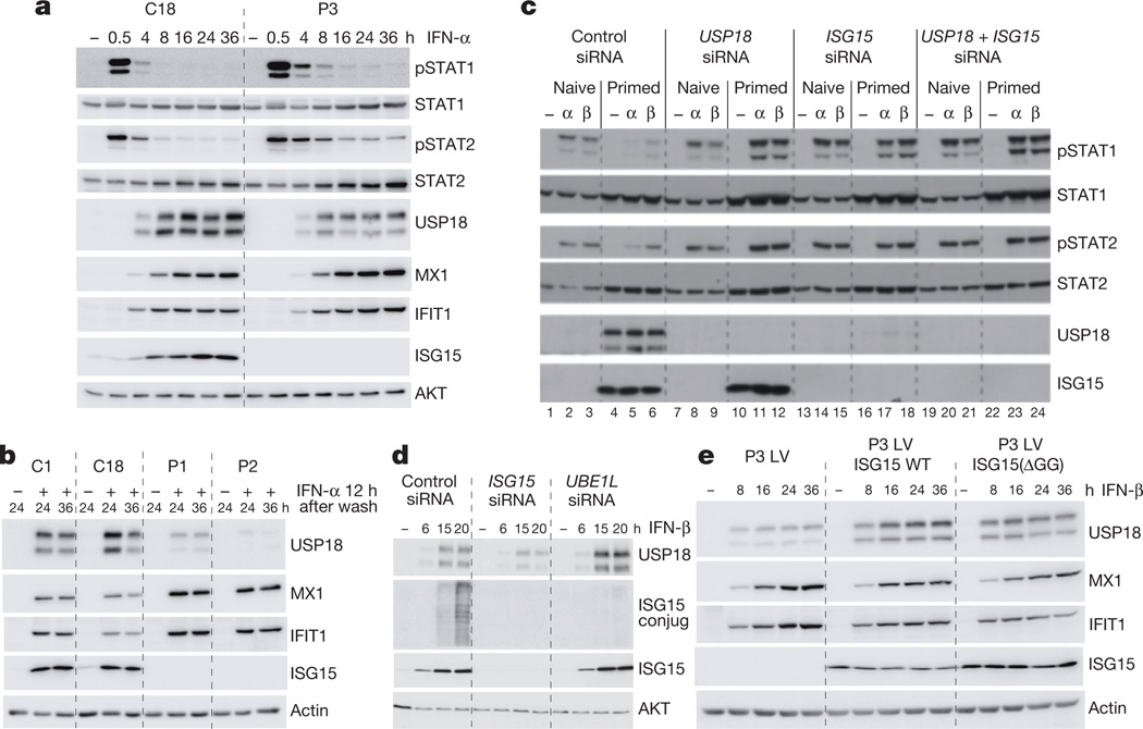
Figure 3.
a, hTert-immortalized fibroblasts from a control (C18) and patient P3 were treated with 100pM IFN-α2 for 0.5 to 36 h. Cell lysates were analysed by western blot for levels of phosphorylated STAT (pSTAT) proteins and proteins encoded by interferon-stimulated genes. b, hTert-immortalized fibroblasts from controls (C1, C18) and patients P1 and P2 were treated with 100pM IFN-α2 for 12 h, washed and left to rest for 24 or 36 h. Protein levels were assessed by western blot. c, HLLR1-1.4 cells were transfected with control short interfering RNA (siRNA) or with siRNA targeting USP18, ISG15 or both. One day later, cells were left untreated (naive) or were primed for 8 h with 500 pM IFN-β, washed and left to rest for 16 h, and then restimulated for 30 min with 100 pM IFN-α2or IFN-β. Lysates were analysed with the indicated antibodies. d, HLLR1-1.4 cells were transfected with control siRNA, ISG15 siRNA or UBE1L siRNA. One day later, IFN-β (500 pM) was added for various periods of time. Lysates were analysed as indicated. e, hTert-immortalized fibroblasts from P3 transduced with lentiviral particles expressing RFP and luciferase (LV), wild-type ISG15 (LV ISG15 WT) or the IS15(ΔGG) mutant (LV ISG15(ΔGG)) were stimulated with IFN-β (500 pM) for 8 to 36 h and protein levels assessed by western blot.

