Fig. 5.
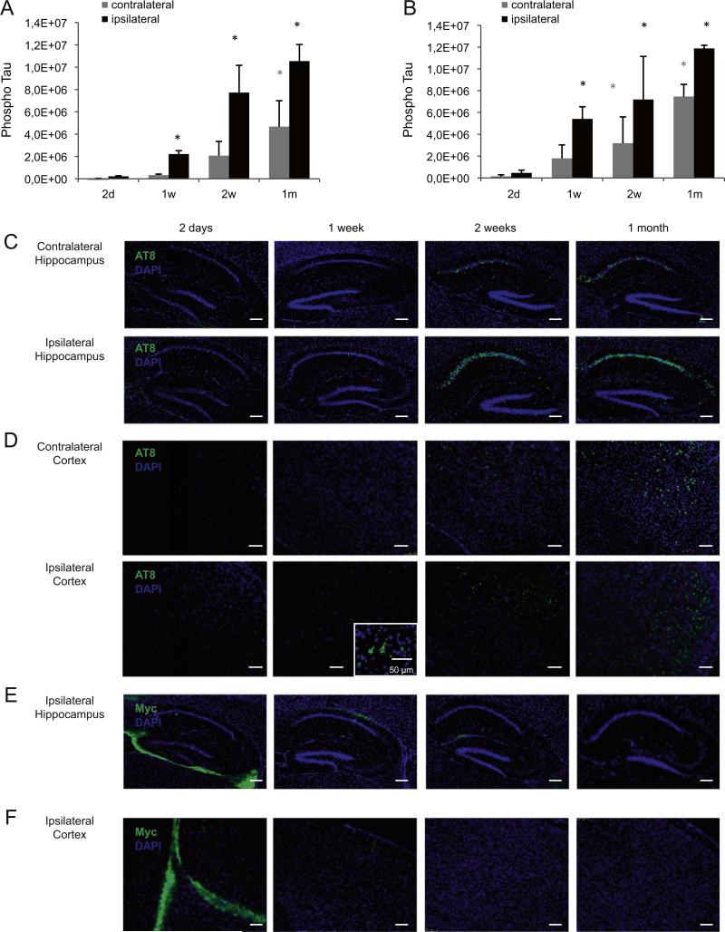
Early onset of tau pathology in mice injected with tau PFFs (A–B) Sarkosyl insoluble brain fractions of the injected and contralateral hemisphere were analyzed for phosphorylated tau at different time points (2 days–1 week) after injection of aggregated K18-PL (25 μg) in the hippocampus (A) or frontal cortex (B). Tau pathology is first observed in the injected hemisphere 1 week after injection and increases up to 1 m after injection in both hippocampus and cortex injected mice. The contralateral hemisphere shows a lower amount of tau phosphorylation and a later onset of pathology. One-way Anova *p < 0.05 vs 2 days (n = 5/group). (C–D) AT8 immunohistochemisty in the hippocampus and frontal cortex of the injected (L +2 mm) and contralateral (L −2 mm) hemisphere confirms the biochemistry findings in animals injected with K18-PL in the hippocampus (C) and frontal cortex (D). (E–F) Myc labeling at the level of injection (L +2 mm) reveals a clear signal around the injection site 2 days after K18-PL injection in hippocampus (E) or frontal cortex (F). This signal decreases 1 and 2 weeks after injection and completely disappears 1 m post-injection. Scale bars: 250 μm.

