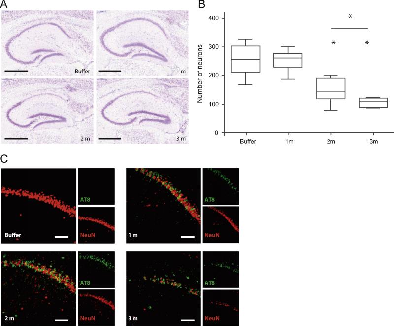
Fig. 8.
Injection of tau PFFs induces cell loss in the hippocampus. (A) Nissl staining in the hippocampus of P301L mice injected with buffer or aggregated K18-PL (25 μg) in the hippocampus. Scale bar: 500 μm. (B) Quantification of the number of Nissl-positive neurons in the CA1 region indicates a clear reduction in the number of neurons 2 and 3 m after K18-PL injection when compared to buffer injected mice. One-way Anova *p < 0.05 vs buffer, #p < 0.05 vs 2 m (n = 53 m buffer, n = 101 m en 3 m K18-PL, n = 82 m K18-PL). (C) AT8-NeuN double labeling in the hippocampus of K18-PL injected animals reveals a decrease in NeuN staining with increasing time after K18-PL injection. AT8-positive neurons are apparent 1 and 2 m after K18-PL injection, but this number is reduced 3 m after injection. Scale bars: 100 μm.

