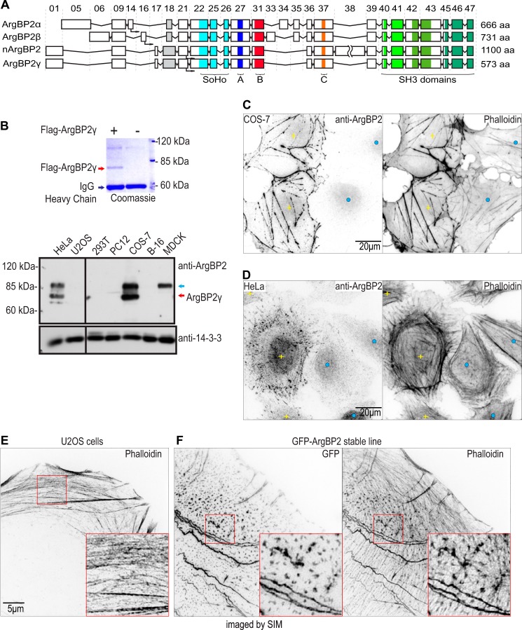
FIGURE 1.
ArgBP2 localizes to actin stress fibers. A, schematic exon map of ArgBP2α, ArgBP2β, nArgBP2, and ArgBP2γ. Exon numbers are as indicated. The sorbin homology and SH3 domains, and conserved regions denoted A, B, and C as described in this study are marked accordingly. B, Western analysis of ArgBP2 expression in various cell lines. FLAG-ArgBP2γ was immunoprecipitated and run on 9% SDS-PAGE, then stained with Coomassie Blue to determine its mobility (top of panel). ArgBP2γ (red arrow) and ArgBP2α/β (blue arrow) were detected in HeLa and COS-7, whereas MDCK cell express only ArgBP2α/β. Typical staining patterns of F-actin and ArgBP2 in COS-7 cells (C) and HeLa cells (D) with detectable (yellow plus) or undetectable (blue dot) ArgBP2 expression are shown. Cells that are ArgBP2 positive cells present with thicker actin stress fibers. ArgBP2 is localized in distinct puncta along the dorsal and ventral actin stress fibers in HeLa cells. The fluorescent signal was inverted (white to black) for greater clarity. E and F, control U2OS cell (E) and GFP-ArgBP2γ localization (F) in U2-OS imaged by SIM. Distribution of the GFP-tagged protein closely resembles that of endogenous ArgBP2 in HeLa cells. GFP-ArgBP2γ expressing cells are characterized by denser and more cross-linked actin stress fibers. The red box marks the region enlarged to highlight the increased bundling at the puncta. The fluorescent signal is inverted (white to black) for greater clarity.

